

Tools at the Lab
Zeiss 880 Confocal Microscope integrated with full Incubation system and an Airyscan Detector
An optical imaging technique for increasing optical resolution and contrast of an object, while capturing multiple 2D images ("slices") at different depths in a sample which enables the reconstruction of 3D structures within an object.
Left: the microscope. Top right: zoom-in on the lense and the light condenser. Bottom right: zoom-in on the control panel of the stage and the monitoring screen.

Chiaro Nano-Indentation System (from Optics11)
Nanoindentation instrument to explore the micro-mechanical properties of cells, spheroids or other small samples. Used to measure forces on cells and micro-materials while placed on an inverted microscope (Left). Right: zoom-in on the probe which is connected to high-precision XY-stage.


Hybrid Rheometer
A rotational and shear type rheometer used to measure the viscosity of a gel in response to applied forces. Left: the rheometer.
Top right: color display, reports a variety of real-time data to the test station to facilitate sample loading, and provides system information during experiments. Bottom right: temperature control system.
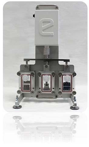
EBERS TC-3 Load Bioreactor
This device allows to mechanically stimulate culture substrates and scaffolds by the application of direct tension and compression mechanical load.

SENSAPEX Micromanipulator
A device used to physically interact with a sample under a microscope, where a level of precision of movement is necessary that cannot be achieved by the unaided human hand. Left: the micromanipulator which is placed on the microscope stage. Right: the control panel and the monitoring screen.

Digital Volume Correlation Algorithm
Digital volume correlation (DVC) is a novel technique for full 3D strain and deformation measurements. The technique imports volume images of the component in reference and deformed states (using confocal microscopy) and is able to calculate the full 3D displacement and strain map by tracking the translations of some pattern cursors.
Right: time-series confocal images of a needle that penetrates into agar gel. Left: 3D displacements field computed by the algorithm.
Finite Element Modelling
A general numerical method for obtaining an approximate solution of a physical problem. It is accomplished by solving partial differential equations in two or three space variables. To solve a problem, the FEM subdivides a large system into smaller, simpler parts that are called finite elements.
Modeling cells embedded within a fibrous gel. Imaging of two green fluorescent protein (GFP)-labeled fibroblasts embedded in fibrin gel (Left) that was imaged by confocal microscopy, compared with a Finite-Element model of two contracting cells in fibrous network (Middle). Applying contraction to the cells resulted in high-stress fibers in the region between the cells (Right).
